NASA’s Curiosity rover has found spectral evidence of an iron-oxide mineral called hematite (Fe2O3) on a rock near Mount Sharp in Gale Crater, Mars.
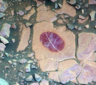
In an area on Mars’ ‘Vera Rubin Ridge’ where the Curiosity team sought to determine whether dust coatings are hiding rocks’ hematite content, the rover found a promising target — a rock called ‘Christmas Cove.’
On September 16, 2017, during the 1,118th Martian day of Curiosity’s work on the planet, the rover’s wire-bristled brush — the Dust Removal Tool — brushed an area about 2.5 inches (6 cm) across.
Curiosity’s Mars Hand Lens Imager (MAHLI) took an image of the freshly brushed area later the same day.
“Removing dust from part of the Christmas Cove target was part of an experiment to check whether dust is subduing the apparent indications of hematite in some of the area’s bedrock,” the rover-team researchers explained.
“The brushed area’s purplish tint in an image from the MAHLI was characteristic of fine-grained hematite, an iron-oxide mineral that can provide information about ancient environmental conditions.”
“Brushing of this target also exposed details in the fine layering and bright veins within the bedrock of this part of Vera Rubin Ridge.”
Curiosity’s ChemCam instrument examined an area on the Christmas Cove and found spectral evidence of hematite. The upper-left inset of this graphic is an image from ChemCam’s Remote Micro-Imager with five labeled points that the instrument analyzed. The image covers an area about 2 inches (5 cm) wide, and the bright lines are fractures in the rock filled with calcium sulfate minerals. The five charted lines of the graphic correspond to those five points and show the spectrometer measurements of brightness at thousands of different wavelengths, from 400 nm (at the violet end of the visible-light spectrum) to 840 nm (in near-infrared). Sections of the spectrum measurements that are helpful for identifying hematite are annotated. These include a dip around 535 nm, the green-light portion of the spectrum at which fine-grained hematite tends to absorb more light and reflect less compared to other parts of the spectrum. The spectra also show maximum reflectance values near 750 nm, followed by a steep decrease in the spectral slope toward 840 nm, both of which are consistent with hematite. Image credit: CNRS.
The next day — September 17, 2017 — observations with the rover’s Mast Camera (Mastcam) and its Chemistry and Camera (ChemCam) confirmed a strong presence of hematite.
“ChemCam sometimes zaps rocks with a laser, but can also be used, as in this case, in a ‘passive’ mode,” the scientists said.
“In this type of investigation, the instrument’s telescope delivers to spectrometers the sunlight reflected from a small target point.”
Source: Sci-news


No comments:
Post a Comment